
Contents
What is Infantigo?
Infantigo is a bacterial infection of the superficial skin, although complicated cases may present with deep skin and fascia involvement as well. It is typically a disease of infancy and childhood; however it may occur in adults too.
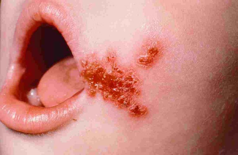
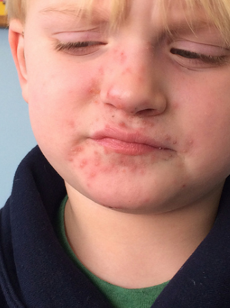
It is usually identified by its typical honey-colored or yellowish crusts on the face (particularly around the child’s nose and mouth), arms, or legs which may or may not be preceded by red colored sores.
Less common presentations of this disease are the formation of large blisters on the groin or armpits. These sores, crusts or blisters maybe painful or itchy, however fever is uncommon in infantigo.
Is infantigo and impetigo the same thing?
Yes Infantigo and impetigo are the same medical terms for one disease
Other names for infantigo include: infintigo, infatigo, infentigo, enfantago, infantago, infitigo, infintago, impetigo or school sores.
What causes infantigo? What are the risk factors for developing infantigo?
The most common bacterial cause for infantigo are the gram positive cocci, Staphylococcus aureus or Streptococcus pyogenes.
Although our skin comes into contact with a number of microorganisms each day, we do not always develop infantigo.
In fact it is only when the continuity of the skin is broken down that these bacteria gain entry into the skin and multiplies over there to cause infection. These risk factors are:
- Mosquito bites
- Scabies
- Herpes
- Eczema
Other risk factors for developing this disease include:
- Overcrowding: Overcrowding is a general risk factor for all contagious diseases as the probability of transmission increases.
- Poor nutrition: Poor nutrition leads to weakened immune defenses of our body and hence poor defense against diseases.
- Age: It is more common in children than in adults.
- Diabetes: Diabetes leads to weakened immunity as the bactericidal function of phagocytes and other white blood cells are severely hampered.
- Contact sports: Contact during sports aids in the transmission of bacteria from an infected person to a non-infected person.
- Daycare: Two risk factors combine in daycares. First is age. Second is overcrowding and hence increased risk of transmission.
How many types of infantigo are there?
There are three types of infantigo. The major types include the bullous and non-bullous infantigo and the less common type is called ecthyma.
- Non-Bullous infantigo: Non-bullous infantigo, also referred to as infantigo contagiosa or contagious infantigo is the most common type of infantigo. It usually starts as a red sore around the mouth or nose. These are usually painless but may cause irritation, discomfort and cosmetic hindrance. It then breaks leaking fluid which dries to form a yellowish or honey colored scab. It then resolves without leaving a scar. During the active stage of disease, regional lymph nodes maybe palpable but fever is absent in most cases.
- Bullous infantigo: It is the second most common type of infantigo. It is mainly seen in children whose age is less than 2 years and is characterized by painless blisters which are filled with fluid and are present on the entire body particularly arms, legs and trunk. The blisters are surrounded by a ring of red skin due to inflammation of the surrounding area which is not tender. The blisters vary in size from a few millimeters to few centimeters. When these blisters rupture, the fluid oozes out and when it dries on exposure to air forms a yellowish or honey-colored scab.
- Ecthyma: The least common type of infantigo is characterized by painful sores which are filled either by clear fluid or by pus. These sores then become ulcers which involve the deeper layers of skin and this is what causes pain. Like other types of infantigo, when the break open, they for scabs. However scars are left behind after the resolution of this type of infantigo hence this is the most cosmetically dangerous type. Regional lymph nodes are usually palpable in this sub-type.
Is infantigo contagious?
Infantigo is highly contagious. The most common mode of transmission is through direct contact with the person who has already been infected. Touching the lesion is almost a guarantee of getting infantigo.
Indirect transmission through contact with fomites such as bed sheets, towels, toys which have been used by infected individuals is another mode of transmission although this mode is less common in causing disease than direct person-to-person transmission.
Another form of transmission which is overlooked but is important is the transmission of disease from one part of the body to another. And this happens when he scratches a lesion when it becomes itchy and then later touches another body part causing implantation and infection of that region of the skin.
How long is infantigo contagious?
After e the medicine is taken, for least 24-48 hours, the impetigo isn’t contagious anymore. In regular cases, after 3 days, the sores should begin to heal.
How long infantigo lasts? How long bullous infantigo lasts?
Nonbullous type of inafantigo usually lasts for 2 weeks (with no scarring). Bullous impetigo typically last much longer (more than a month). Both respond well to treatment.
Infantigo incubation period
The incubation period for staphylococcus aureus is 1 to 3 days and for streptococcous pyogenes is 10 days. This means that if the infected individual is infected with staph aureus, then the exposed individual will develop signs and symptoms within 1 to 3 days and if the infected individual is infected with strep pyogenes, then the exposed individual will develop signs and symptoms within 10 days.
However if he does not develop any of the aforementioned signs and symptoms, then most probably he will not develop it at all unless exposed for a second time or to a second person.
How to prevent the spread of infantigo?
How long does a person continue to spread infantigo after he has become infected?
As explained above, direct transmission is the most common mode of transmission and all patients should take measures to stop this transmission as the complications of this disease are fatal.
One of the best ways to prevent any skin disease is to keep your skin clean. Therefore it is recommended to wash cuts, wounds, scrapes or insect bites by an antiseptic solution or gentle flowing clean tap water. If the wounds are clearly contaminated one should consult the ER department immediately.
If you have diagnosed infantigo, then take the following measures to help spread it to others:
- Wash the infected areas/lesions with mild soap and then gentle flowing tap water or with an antiseptic solution and then cover it with wire gauze and dressing.
- The infected person’s towels, clothes and lines should be washed every day and be kept separate so that no other family members share them.
- It is advised to wear soft gloves when applying the antibiotic cream. After applying one should remove the gloves and then thoroughly wash his or her hands.
- A child’s nail is a rich reservoir for these gram positive cocci. So his nails should be cut so that this reservoir is never built up which might infect other children. Nail cutting also helps in preventing damage which is associated with scratching.
- Needless to say, wash your hands frequently particularly after scratching.
- Infantigo is a contagious disease. So isolation from susceptible individuals is another useful approach. Keeping this in mind, all parents should keep their children at home so prevent other children from getting this disease.
In areas where mosquito bites are very common, people should be advised to sleep under nets or apply mosquito repellant lotions especially when there is an epidemic of infantigo.
Infantigo diagnosis
Infantigo is usually diagnosed clinically i.e. on the basis of history and signs and symptoms. However in areas where methicillin resistant staphylococcus aureus (MRSA) is common, bacterial culture and sensitivity tests are recommended to identify them. Other indications for these tests are if there is an outbreak of infantigo in a day care, family or athletic team settings or if post streptococcal glomerulonephritis is present.
In suspected cases of acute post streptococcal glomerulonephritis (APSGN), evidence of previous streptococcal skin infection should be asked.
The laboratory specimen in bullous lesion is the blister fluid and in non-bullous lesions is the fresh exudate which can be obtained by cleansing the crusts and then uplifting the scab. The tests normally performed on these specimens are the bacterial culture and sensitivity tests and gram stain.
If you see gram positive cocci arranged in chains, it indicates the presence of streptococcus pyogenens and if you see gram positive cocci arranged in clusters, it indicates the presence of staph aureus. Culture and sensitivity tests help the physician to decide proper antibiotic therapy.
If your patient is a suspect of ASPGN, then history of a recent streptococcal skin infection should be asked. Apart from this, obtaining titers of antideoxyribonuclease B (anti-DNase B) and antihyaluronidase (AH) antibodies can prove to be helpful in confirming your diagnosis as more than 92% patients of infantigo associated acute post streptococcal glomerulonephritis have elevated anti-DNAse B titres.
Also note that testing for antistreptolysin O (ASO) antibodies is a poor test for this purpose as only 51% with infantigo associated acute post streptococcal glomerulonephritis have elevated ASO titres. Urinalysis is the final test used for confirming your diagnosis. A patient who has recently developed edema or hypertension post skin infection is an ideal candidate for undergoing this test. Hematuria, proteinuria, and cylindruria indicate renal involvement.
A patient with recurrent infantigo or a patient who has no obvious exposure to infantigo may undergo a bacterial culture of the nares as sometimes staph aureus becomes a resident of nasal mucosa causing recurrent infantigo. If his nasal passages are clear and he is still suffering from persistent recurrent episodes of infantigo, bacterial cultures should be obtained from the pharynx, axillae, and perineum.
In case tests from the above mentioned locations give a negative staph aureus carrier status and there are no obvious predisposing factors such as a preexisting dermatosis, one should obtain serum IgM IgA, IgG, including IgG subclasses, as then the patient might be suffering from an immunocompromised state which is going undetected.
Histological Picture
Biopsy is indicated in either doubtful cases or in recurrent cases of infantigo. Bullous infantigo is characterized by very few or no inflammatory cells within the bulla. Polymorphonuclear cells are present in the upper dermis and acantholysis (loss of intercellular connections) is seen in the granular layer of skin.
Non-bullous infantigo is characterized by a serum crust covering the epidermis from the outside. Within this crust, neutrophils and gram positive cocci may be seen. Intercellular edema in the epidermis is seen. This is called epidermal spongiosis. There is infiltration of neutrophils and lymphocytes in the epidermis as well.
Infantigo differential diagnosis
Tinea is a common disease of the skin and oftens mimic imfantigo. However close examination will reveal the difference as tinea almost never occupies the upper lip.
If the lesion does not show a typical clinical picture, then either a potassium hydroxide wet mount or a tzanck preparation & viral culture maybe performed. These tests help to rule out bullous dermatophyte infection and herpes simplex infection respectively. There is a documented high risk of patients with eczema developing herpetic infantigo.
Follicular mucinosis is another disease similar to infantigo in clinical presentation. Although follicular mucinosis often does not have a crust, differentiating these two diseases in the facial region is very difficult and often needs a biopsy.
Erysipelas is a skin infection which not only resembles infantigo but is also caued by the same bacteria; group A beta-hemolytic streptococci. These two conditions can be differentiated clinically as erysipelas manifests as a sharply demarcated erythematous plaque caused by dermal inflammation
Other diseases to be considered in the differential of non-bullous infantigo are:
- Cutaneous candidiasis
- Kerion
- Inflammatory dermatophytosis
- Dermatophytic Infections
- Discoid lupus erythematosus
- Sweet syndrome (acute febrile neutrophilic dermatosis)
Other diseases to be considered in the differential of bullous infantigo are:
- Linear immunoglobulin A bullous dermatosis
- Bullous pemphigoid reactions
- Bullous lupus erythematosus
- Bullous scabies
- Dermatitis herpetiformis
- Bullous-fixed drug reaction
Infantigo treatment
Infantigo will usually resolve within two weeks after the appearance of symptoms, however since few cases do develop complications it is always advisable to get treatment for it.
The approach for treating infantigo revolves around local wound care with antibiotic therapy. Either topical antibiotics alone are used or a combination of both topical and systemic antibiotics is used.
Infantigo wound care
Cleansing of wound and removal of crusts using mild antibacterial soap and gentle flowing tap water or antiseptic solution followed by dressing of the lesion is recommended. Chlorhexideine or sodium hypochlorite baths are said to prevent transmission and recurrences however the effectiveness of this has not been proven to date.
Antibiotic therapy for infantigo
As mentioned above, almost all cases of infantigo are due to infection with two important gram positive cocci. These are Staphylococcus aureus and Streptococcus pyogenes. Therefore the antibiotic used for treating infantigo must provide cover against these two organisms.
It is noteworthy that since the prevalence of methicillin-resistant S aureus (MRSA) and erythromycin-resistant Streptococcus is on the rise, infantigo might not respond to methicillin or to macrolides. The prevalence of MRSA is so high that it was found to be responsible for 78% of all community acquired staphylococcal-related skin and soft tissue skin infections in a multicenter US study.
Community-acquired MRSA (CA-MRSA) is and more likely to cause suppurative infections such as abscesses and less likely to cause exfoliative infections such as infantigo because it possesses the Panton-Valentine leukocidin gene rather than the exfoliative toxin gene.
So while minor skin and soft-tissue infections may be empirically treated with, first-generation or second-generation oral cephalosporins, macrolides, semisynthetic penicillin or clindamycin, major or unresponsive cases must be given trimethoprim/sulfamethoxazole or doxycycline (the preferred tetracycline for skin infections is doxycycline although other tetracyclines may also be given) in children older than 8 years.
The problem with using doxycycline or minocycline is that failure rates are as high as 21% with these drugs; therefore the physician must re-evaluate his patient after 48hours to check the response of these drugs.
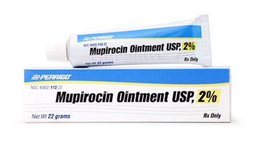
Single lesions of nonbullous infantigo or small areas of involvement usually resolve with topical mupirocin or retapamulin. Systemic antibiotics are indicated for nonbullous infantigo with extensive involvement, childcare clusters, multiple family members, in athletic teams, or for patients suffering for bullous infantigo.
Patients suffering with bullous infantigo involving large areas of the body with ruptuted bullae and denuded skin have lost a tremendous amount of fluid. As a result their management also involves intravenous fluid resuscitation which is given at a volume and rate similar to standard volume replacement for burns.
Hospital admissions are warranted for those cases of infantigo who either have extensive disease or for infants at risk of sepsis and/or dehydration due to skin loss or in a child with untreated infantigo. Hospital admission provides the benefit of contact isolation.
Topical antibiotic treatment for infantigo
Topical therapy is the treatment of choice for uncomplicated localized infantigo. It eradicates disease and reduces the risk of disease transmission. It is easier to use and can be used without any worry of systemic side effects.
Disadvantages of this mode of treatment include the inability to remove these bacteria from respiratory tract (these bacteria if left untreated may cause recurrence) and difficulty faced in applying the antibiotic cream to extensive lesions and lesions on obscure areas. The three important topical antibiotics for treating infantigo are mupirocin, retapamulin and fusidic acid.
Mupirocin ointment is used for treating lesions as well as to clear nasal carriers. It is equally effective as oral cephalexin. The standard course is of 5 days, although the verification of this duration has still not been done in large studies. Recently MRSA resistance to this drug has emerged.
Retapamulin is a relatively new drug. It is FDA approved for treating localized infantigo. It is found to have activity against even those gram positive cocci that are resistant to multiple antibiotic drugs, such as methicillin, erythromycin, fusidic acid, mupirocin, azithromycin, and levofloxacin. It is also founf to have activity against erythromycin-resistant S pyogenes, fusidic acid–resistant and mupirocin-resistant S aureus, and MRSA (including P-VL–positive strains).
Topical sodium fusidate is another topical antiobiotic which is used for localized infantigo, however due to its high resistance rates; it is no longer in use in the United States.
Systemic antibiotic treatment for infantigo
Widespread or complicated infections or those infections which are associated with systemic manifestations need systemic antibiotic treatment. Systemic therapy is also indicated when there are multiple incidents of pyoderma in a day care, family or athletic team settings.
Cephalexin is the drug of choice for oral antibacterial therapy in children. Other antiobiotics commonly used include the Beta-lactamase‒resistant antibiotics (eg, cephalosporins, amoxicillin-clavulanate, dicloxacillin. A common problem is the infection by Community-acquired MRSA.
In such cases alternate drugs such as trimethoprim/sulfamethoxazole, and doxycycline maybe used in patients older than 8 years. Clindamycin was another alternate but its use for treating MRSA is becoming limited as it has been discovered that MRSA strains can also develop resistance to clindamycin.
Erythromycin maybe given to patients with penicillin hypersensitivity, however since its resistance is also on the rise, geographical knowledge about where these strains are more common is a must have. In such areas clindamycin maybe used.
Complications of infantigo
Infantigo usually self resolves within a couple of weeks and even earlier with the use of antibiotics. Therefore its complications are very rare and are seen in less than 2 percent of the population suffering from infantigo. The complications are:
- cellulitis
- scarlet fever
- guttate psoriasis
- septicemia (a type of sepsis)
- post-streptococcal glomerulonephritis
Cellulitis as a complication of infantigo
Cellulitis is the bacterial infection of the deeper layers of the skin, particularly dermis and the subcutaneous fat. Although isolation of bacteria from the lesions is difficult, but the appearance of cellulitis after infantigo and response to penicillin suggest that the cause is a streptococcal bacteria.
The signs and symptoms of cellulitis are non-specific and hence it may be confused with other infective lesions of the skin. There is an area of redness whose borders are not well defined and which may increase in size if left untreated.
The area around the red skin is usually swollen. This area of redness may turn white on applying pressure. Fever, fatigue, malaise, headache and involvement of lymphatic vessels may also be seen. All these symptoms are almost always present with pain.
The mainstay in the treatment of cellulitis is the antibiotic flucloxacillin which is penicillin. However flouroquinolones may also be used. Less notorious analgesics such as paracetamol are usually sufficient to overcome the pain and fever associated with cellulitis. Hospital stay will be indicated in those individuals who cannot take good care of themselves at home or who have severe infection.
Guttate Psoriasis as a complication of infantigo
Guttate psoriasis or eruptive psoriasis is type of psoriasis characterized by small lesions over the proximal extremities and upper trunk. The size of these lesions rarely exceed 1.5cm in diameter. These lesions have a drop-like appearance and hence it is called guttate which is Latin for drops.
The usual trigger for this type of psoriasis is a strep throat; however some cases have been linked to infantigo as well. Initially red spots appear which are dry and itchy. When these spots are scratched, the dry skin over them is removed leaving behind red skin with white, dry areas marking where flakes of dry skin stop and start. Some spots may have a pale yellowish area in the center.
Knowledge of the appearance of these spots is importance as the diagnosis of guttate psoriasis is usually done by clinical examination alone as a skin biopsy cannot be done in every suspected case.
Since there are no form guidelines for treating guttate psoriasis, its treatment is similar to treatment of plaque psoriasis:
- Cortisone cream to help stop itching and swelling
- Dandruff shampoo for your scalp
- Lotions with coal tarto soothe your skin
- Moisturizers
- Prescription medicines with vitamin D or vitamin A
If the physician deems your case more serious, then he might prescribe the following drugs:
- Corticosteroids
- Methotrexate
- Cyclosporine
Systemic antibiotics are also prescribed sometimes, however their efficacy in treating post infantigo guttate psoriasis has been studied till now.
Scarlet fever as a complication of infantigo
Scarlet fever is an infectious disease caused by group A streptococcus that is characterized by a fine, red rash across the body. The rash feels like a sandpaper and due to this the tongue may be red and bumpy, an appearance called ‘strawberry tongue’. Other common signs and symptoms of the disease include sore throat, fever, headaches and swollen lymph nodes
The diagnosis of scarlet fever depends upon clinical signs and symptoms and lab tests. CBC shows marked increase in white blood cell count with neutrophilia and conserved or increased eosinophils. Erythrocyte sedimentation rate is elevated and so is C-reactive protein. Titer for antistreptolysin O is also high. Throat culture or skin culture will be positive but blood culture is usually negative.
The most common treatment for scarlet fever is a 10-day course of penicillin (or erythromycin for those who are allergic to penicillin). Paracetamol is given to control fever and pain.
An important point in the management of scarlet fever includes isolating the child which has been diagnosed with it because it is highly contagious and might prove to be fatal if left undiagnosed.
Septicemia as a complication of infantigo
Septicemia is infection of the blood. It occurs when a bacterial infection of some other part of the body (in this case the skin) enters the bloodstream. This will cause dissemination of bacteria and their toxins throughout the body and cause sepsis.
Therefore it is essential that if you were suffering from infantigo and now develop chills, fever, tachycardia or an increased respiratory rate. If left untreated septicemia will be converted to sepsis which is characterized by more severe signs and symptoms such as nausea and vomiting, confusion, red dots on skin, decreased urine output and shock.
Diagnosing septicemia will depend upon medical history, clinical examination and lab tests. Simple measurements such as body temperature, heart rate and breathing rate will be helpful in diagnosis. Blood culture will also be positive; the most likely organism in such a case would obviously be Staphylococcus aureus or Streptococcus pyogenes. A wound culture may also be done.
The treatment for sepsis depends upon the severity of signs and symptoms. If it is detected early and no damage to vital organs is found, then it may be possible to send the patient home after prescribing them antibiotics.
Almost everyone who consults a doctor at this stage makes a full recovery. Hospital admission is warranted in individuals with severe sepsis and septic shock. Some of them may need admission to an intensive care unit (ICU). This is because patients who present late with sever sepsis have compromised function of vital organs and need support to make a full recovery.
Acute Poststreptococcal Glomerulonephritis (APSGN) as a complication of infantigo
Acute poststreptococcal glomerulonephritis is a disorder of the small blood vessels of kidney called glomeruli after infection of throat or skin by streptococci. It is characterized by hematuria (cola colored urine), proteinuria, red blood cell casts in the urine, edema, and hypertension with or without oliguria. It occurs 3-6 weeks after infantigo and 1-2 after throat infection.
Diagnosis of APSGN is primarily based on lab tests which focus on providing evidence of previous streptococcal infection, renal function studies and serologic studies. Biopsy may also be warranted in some cases. Previous streptococcal infection can be found out using anti–DNAse B and AHase titers which are usually positive following skin infections.
Elevated serum creatinine values and blood urea nitrogen (BUN) and are indicative of renal involvement and a decrease in the glomerular filtration rate that occurs in the acute phase. Serologic findings include low serum complement levels due to antigen-antibody interaction. Biopsy shows the following features:
There is hyper cellularity of glomeruli due to presence of endothelial and mesangial cells and migrant inflammatory cells
On immunofluorescence, a diffuse granular pattern is present along the glomerular capillary wall and mesangium due to deposition of immunoglobulin G and C3.
Electron microscopy reveals thickening of glomerular basement membrane. The most consistent feature is the presence of humps due to subepithelial deposition of electron-dense immune-type deposits and these are found on the part of the glomerular basement membrane overlying the mesangium
Treatment of APSGN includes symptomatic therapy against edema and blood pressure. The medicine and dose prescribed depends upon clinical severity of the illness. First course of action is to restrict salt and water intake. In addition diuretics, particularly loop diuretics are prescribed due to their rapid onset of action.
Other drugs which can be used for controlling hypertension and edema include calcium channel blockers, angiotensin-converting enzyme inhibitors (ACEIs), or angiotensin receptor blockers (ARBs). However ACEIs and ARBs can cause hyperkalemia which temporarily halts recovery of renal function.
Specific antibiotic therapy against streptococci is an integral part in treating APSGN. The preferred course is a 7 or 10 day course of penicillin or erythromycin if you develop anaphylaxis with penicillin.
Home remedies for Infantigo
White Vinegar for infantigo
White vinegar keeps the affected area clean therefore promoting healing of the skin sores. Mixing 1 tablespoon of white distilled vinegar in 2 cups of lukewarm water will make your own antibiotic solution. Then use a cotton ball to wash the infected skin with this solution before applying your prescribed antibiotic treatment. Repeat 2 to 3 times daily.
Heat therapy for infantigo
Heating will help kill the bacteria as well as help dry the sores and lesions. You can do this by soaking a cloth in hot water and then wring out the excess liquid. Apply this cloth to the lesion. Repeat this 3-4 times a day.
A blow dryer can also be used to apply heat to the affected area.
Tea tree oil for infantigo
Tea tree oil has both antimicrobial and anti-inflammatory properties that can halt the spread of this bacterial infection. Few drops of tea tree oil added to a small tub of lukewarm water yields a solution that can be used to wash the affected area a few times daily.
Grapefruit seed extract for infantigo
Grapefruit see extract is has antibacterial and antioxidant properties so it works as a non-toxic disinfectant. It can be taken in the form of a supplement. However if you want to avoid taking any supplements, a homemade solution can be made by adding a few drops of extract to 2 tablespoons of water. This solution can then be applied on the lesion using a cotton ball.
Garlic for infantigo
Garlic is known to be used in the treatment of over 100 diseases, one of these is infantigo. Garlic is one of the most potent natural antibiotics. A solution for topical use can be obtained by heating 2 tablespoons of sesame oil and frying 2 or 3 minced garlic cloves in it. Then cool and strain the oil. Apply this solution on the affected area once or twice daily for a few days.
Manuka honey for infantigo
Antibacterial, antiseptic and healing properties of manuka honey inhibit the growth of the bacteria. It also boosts your immunity increasing the speed of the healing process. Ideally some manuka honey should be applied on the affected area and then left for a few hours before washing it off with lukewarm water. Repeat a few times daily for a few days. Also eating 1 to 2 teaspoons of manuka honey daily boosts our immunity and helps the body fight the bacteria.
Goldenseal for infantigo
Lovers of herbs can use goldenseal for treating infantigo which can disinfect the skin sores and blisters and also strengthens our immune system. There are 3 ways this herb can be used:
- 1 teaspoon of goldenseal powder is mixed in 1 cup of hot water for 10 minutes. This herbal tea is then allowed to cool. Clean the infected area twice daily with this tea.
- Goldenseal supplements are available and can be used orally.
- Goldenseal cream or lotion is also available commercially. Apply the cream 2 times daily.
Aloe Vera for infantigo
The antibacterial, anti-inflammatory, antiseptic, antioxidant and skin-repairing properties of aloe vera are well known. The gel extract of a fresh aloe vera is applied directly on the affected skin and left for at least 30 minutes. Reapply 2 or 3 times daily for a week.
Zinc for infantigo
Zinc is a mineral which has been found to play an important role in a number of different body processes as it as a co-factor for many enzymes. It improves immunity by playing role in certain cellular process that counteracts bacterial infections and helps balance the normal immune response. It is also documented that zinc decreases the risk of infantigo among premature babies.
Zinc rich foods such as beans, nuts, some shellfish, whole grains, and fortified cereals are proven to help treat infantigo. If these food products are not readily available then you can use zinc supplements to take 15 to 25mg per day.
Olive oil for infantigo
Olive oil too has antimicrobial properties. The correct way of using olive oil for the treatment of infantigo includes applying a few drops of oil over the lesion and then wait for a few minutes before washing it off. Repeat this exercise a couple of times every day till the lesion resolves.
Turmeric for infantigo
Turmeric is believed to increase our immunity, although there is no study to prove this fact. Nonetheless, mixing powdered turmeric in warm milk and then consuming this mixture twice a day will help treat infantigo at home.
Echinacea for infantigo
Echinacea has antiviral and antibiotic properties. This herb has the unique advantage of preventing dissemination of infantigo. This herb is mixed with water and then the resultant solution is applied over the affected body part. Caution should be practiced in immunosuppressive and AIDS patients since this herb can cause damage in such individuals.
Dietary changes for infantigo
Taking a well-balanced diet will generally improve your health, boost your immunity and protect you from disease. The following dietary changes are recommended:
- Fresh vegetable and citrus fresh juice provide various vitamins and minerals which boost our immunity. They also contain high levels of antioxidants that help the body fight off the infection.
- Fresh apple juice should be taken daily. Two to three glasses would be sufficient.
- Intake of omega-3 fatty acids should be increased.
- Refined sugars and fried foods should be avoided. Although there is no direct evidence linking infantigo with these food products, but since these products are considered to be unhealthy they should be avoided during time of disease.
- Restrict red meat from your diet until the infection has cleared.
- Drinking an ample amount of water to keep your body hydrated is important.
“Scabies – Skin rash, Pictures , Causes, Symptoms, Home Remedy Treatments and Prevention“