
Contents
- What are cysts?
- What is Pilar cyst?
- Can Pilar cyst become tumor or cancer?
- How Pilar cyst looks like?
- What is the prevalence of Pilar cysts?
- What is the difference between Epidermoid and Pilar cysts?
- What causes Pilar cysts?
- What are signs and symptoms of Pilar cysts?
- Pilar cyst histology
- Pilar cyst diagnosis
- Medication treatment for Pilar cyst
- Surgical treatment of Pilar cyst
- Pilar cysts complications
- Breast pilar cysts
What are cysts?
Cysts are sac-like or capsule-like closed structures with a distinct membrane and division compared to the nearby tissue that may be filled with liquid, semisolid material or rarely gaseous material. The unique aspect of a cyst is that the cyst cells are forming the “shell” which is abnormal in the appearance and behavior compared to the surrounding normal tissue.
Genetics, infection and underlying condition are most common causes of cyst forming. Usually cysts are asymptomatic with no evident sign. However, some cysts, especially those of the skin, mucous membranes can be felt as a lump or bump and can be very painful. Some cysts may have symptoms connected to the organs in which they are such as liver, kidneys or pancreas.
Cysts can be diagnosed by palpation, X-rays, ultrasound, CT scans, MRI scans, and needle biopsies. Most cysts do not need to be treated, but in many cases physicians may use needle aspiration or surgical procedures to remove and treat some cysts.
Most cysts can’t be prevented and usually there are no medicines that can reduce or remove cysts. Only cysts caused by infection can be prevented. Cysts prognosis is good, however some types of cysts may have malignant origin.
What is Pilar cyst?
Pilar or Trichilemmal cysts are intradermal or subcutaneous cysts formed from a hair follicle. Other terms for Pilar cysts are: wen, isthmus-catagen cyst or keratin filled cyst. These cysts are very common occurring with incidence of 5-10 % of overall population. In more than 90 % cases they are located on the scalp and are most likely cutaneous.
They are the most prevalent in middle-aged females. Pilar cysts are smooth, usually mobile and filled with protein keratin. The general rule is that Pilar cysts are almost always benign. They can be sporadic or they might be autosomal dominantly inherited so they often run in the family. Pilar cysts may be inflamed and tender, which most commonly depends on whether they have ruptured.
Can Pilar cyst become tumor or cancer?
Rarely, in about 2%, these cysts may have more extensive growth and may form rapidly multiplying pilar or trichilemmal tumors that are also known as proliferating trichilemmal cysts, which are benign but may grow aggressively at the cyst site. Proliferating trichilemmal cysts are progressively enlarging and may be up to 25 cm in diameter. They may form nodules that occasionally ulcerate.
Although they are almost always benign tumors, they may be locally aggressive provoking mechanical issues to the surrounding tissues including blood vessels. Very rarely, trichilemmal cysts can become cancerous. Recurrences and metastases have been also observed, with very rare malignant transformation. There were case reports that have described Merkel cell carcinoma arising from Merkel cells in pilar cysts.
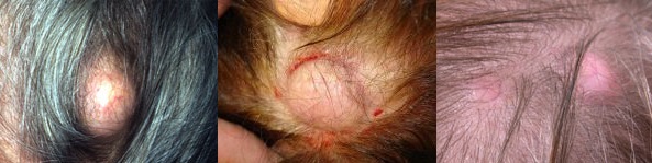
How Pilar cyst looks like?
Pilar cysts are most commonly present as individual, firm, mobile, cutaneous nodules with 0.5 to 5 cm measuring in diameter. As said, they are most commonly found on the scalp. If they are inflamed, they may become painful.
They are generally dome-shaped and flesh-colored. Pilar cysts are fluid-filled cysts. Fluid looks like oily toothpaste. A pilar cyst has a rough wall, but once treated it can be disconnected easily from the skin. They usually grow until they reach a stable size. They don’t burst a lot, mainly because of the thick wall that is not prone to rupture.
What is the prevalence of Pilar cysts?
Pilar cysts are common condition worldwide. It is estimated that pilar cysts occur in 5-10 % of the population. They appear to be in solitary formtion in 30% of cases and multiple in 70% of cases. In 90% of cases they are localized on scalp.
They may be also seen uncommonly on the face, neck, scrotum, trunk, and extremities. Pilar cysts occur more frequently in women than in men, rather in individuals of middle age than in younger persons. Proliferating trichilemmal cysts that may cause tumors are more common in elderly women. There is no known racial predilection.
What is the difference between Epidermoid and Pilar cysts?
In the past, epidermoid and pilar cysts were wrongly diagnosed as ‘sebaceous’ cysts. However, this term still exist and should be used only in situations for much less common type of cysts that are filled with a clear oily liquid made by sebaceous glands.
The main differences between epidermoid and pilar cysts are following:
- Location. Pilar cyst are most commonly localized on scalp (in 90 % of cases), while epidermoid cysts can be mostly found on face, neck and trunk. Pilar cysts are also more commonly found on the scrotum compared to epidermoid cysts.
- Central punctum. Epidermoid cysts have central punctum while pilar cysts don’t.
- Origin. The origin of the pilar cysts is outer root sheath of hair follicle while for epidermal cysts origin is epithelium or hair follicle infundibulum.
- Cyst wall structure. The cyst wall of a pilar cyst is thick and less prone to rupture while epidermoid cyst wall is delicate and prone to rupture.
- Histology. Granular layer is present in epidermoid cysts while it can be found in histology samples of pilar cysts.
- Hereditary. Epidermoid cysts are usually not hereditary, but they may be indirectly caused by conditions that have hereditary origin. Pilar cysts are usually hereditary, being inherited as an autosomal dominant disorder. There is a 50% chance that each child of an affected parent will inherit the condition.
What causes Pilar cysts?
Pilar cysts come from the outer root sheat of the hair follicile. Their true origin is yet unknown, but it has been proposed that they are made as a genetically determined structural aberration. Some theory is that some cells that are normally located nearly to the surface of the skin may get into deeper parts of the skin and continue to multiply.
The cells multiply and form a sac or capsule-like formation producing keratin that they would generally make on the top layer of the skin. The keratin becomes moist and forms a toothpaste-like material.
Different factors such as following skin injury may be the cause since a trauma of the skin can accumulate keratin and infected region will then form thick lumps. Blocked sebacues glands may be also the cause of Pilar cysts. If sebaceous glands are blocked then they cause pilar cysts.
What are signs and symptoms of Pilar cysts?
Most common signs and symptoms of pilar cysts are following:
- Dome-shaped bumps: They grow gradually and look like round bumps under the skin. They are the size of a pea and may grow to be several cm in diameter.
- Pain: Pilar cysts are usually not painful, but it may become if they get infected or ruptured due to mechanical injury. In those cases they usually got very painful on touch.
- Redness: If inflamed or infected they change the color of the skin into red.
- Hair Loss: Localized hair loss is likely to occur at the location where pilar cyst grows.
- Bad Smell: If the pilar cyst get damaged and ruptured, the material they are made of will come out. In such a case, foul smell will come from the infected region.
- Horn: In some cases a horn may appear over the infected region
- Irritation: Irritation is most likely to appear when the pilar cyst is rubbed or scratched against the clothes.
Pilar cyst histology
The histological pathology of a pilar cyst is specific. The cyst wall is stratified with squamous epithelium with a palisaded outer layer, which resembles of outer root sheath of a hair follicle. The inner layer is not characterized with granular layer which is significant indicator for differential diagnosis with epidermoid cysts. The cyst shows very dense pink color of keratin on eosin and haematoxylin staining.
Pilar cyst diagnosis
After physical examination, excision and histopathologic evaluation may be needed to precisely confirm the diagnosis. Rarely, Pilar cysts may be misdiagnosed with Epidermoid cysts. But, pilar cysts are extracted more easily than epidermoid cysts, and this may be enough to confirm the diagnosis.
If a pilar cyst becomes inflamed and get rupture, the cyst should be submitted for histopathology in order to exclude potential carcinoma, especially nodular or nodulocystic basal cell carcinomas. Infected cysts should be cultured in order to precisely be identified what bacteria are source of infection.
Differential diagnosis may be needed for exluding following conditions:
- Acne Keloidalis Nuchae
- Cutaneous Lipomas
- Dermoid Cyst
- Epidermal Inclusion Cyst
- Favre-Racouchot Syndrome (Nodular Elastosis With Cysts and Comedones)
- Pilomatrixoma
- Steatocystoma Multiplex
Medication treatment for Pilar cyst
There are no medications, nor systemic or topical that are able to shrink or resolve a pilar cyst, thus surgical intervention is the only way to get rid of pilar cyst. Antibiotic may be helpful if pilar cyst get infected while corticosteroids injections are effective for inflamed tissue, but these medicines won’t resolve the problem.
Surgical treatment of Pilar cyst
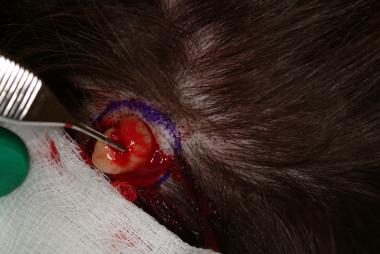
The best treatment option is complete cyst excision. There are several methods that can be used for successful removal of these cysts including:
- Small linear incision
- Elliptical excision or
- Circular dermal punch incision
All of these methods have been prove to be effective ways to remove the cysts.
image : methods to remove cyst
All of these procedures follow next steps:
- Anesthetize the area with local anesthetic.
- A small linear incision, an elliptical excision, or a punch incision should be made over the center of the cyst using a dermal punch biopsy tool
- In most cases, the fibrous capsules of the pilar cysts are so thick that the cyst can be removed at whole through blunt dissection without expression of the cyst material. Alternatively, the contents of the cyst should be expressed, and then, using a curette, pressed against the inner wall, moving it back and forth to dislodge the outer side of the cyst from the nearby tissue.
- The edge of the cyst should be grasped with forceps and the cyst wall should be separated from the nearby connective tissue through blunt dissection.
- Control the bleeding, and suture or dress the wound as necessary.
If the cyst is infected or ruptured, deferring excision until the infection and inflammation is reduced decreases the probability of spreading infection and wound healing problems.
Most proliferating pilar cysts are cured with complete surgical removal. In very cases proliferating trichilemmal cysts require several local excisions.
Pilar cysts complications
- Although most pilar cysts are asymptomatic, cyst infection and rupture may occur.
- Cysts may be traumatized when brushing or combing hair.
- Proliferating trichilemmal may invade surrounding tissue and ulcerate.
- Malignant transformations of pilar cysts are exceedingly rare but can occur
- Very rarely skull lesions after surgery can have an intracranial or intraosseous connection. Inappropriate surgery may result in cerebrospinal fluid leakage and complications including meningitis and death. Therefore, preliminary CT or MRI scan may be of value.
- Surgery will leave a scar. Also, the cyst may reappear, especially if it was not fully removed from the scalp or scrotum
- Anxiety or cosmetic embarrassment
Breast pilar cysts
In the rest of 10% of cases pilar cysts may occur at the different parts of the body including breasts. Pilar cysts are known to be smooth and movable, and usually after finding them with palpitation, women will be concerned about a possible breast cancer.
It should be known that breast pilar cysts are not malign tumors, and very rarely they can develop into tumors which have benign nature. Pilar tumors may show mitotic cells, nuclear atypia, and dyskeratotic cells and this can be a misleading and incorrect indicator of possible skin carcinoma.
On mammogram, a pilar breast tumor may look like a lobulated, well-circumscribed mass. Externally the skin surrounding the tumor may be ulcerated and atrophied.
While they are mostly nonaggressive and benign, they might show local grow, be highly proliferative and even to metastasize. So, the concern would be that while a proliferative pilar tumor is not life-threating, it might start to develop on a critical organ or the brain, and make mechanical issues and serious problems.
Trichilemmomas are usually 1 to 10 cm long in diameter, but, they can really grow sometimes, even reaching sizes up to 25 cm in diameter. Surgical treatment of breast pilar cysts has been shown as a very effective method.
“What causes Dyshidrotic eczema? Is Pompholyx contagious? How do you treat Dyshidrotic eczema?”